Doppler transcraneal

0

ICMPregnant and postpartum women account for a low, although not negligible, rate of admissions to the intensive care unit (ICU), being hypertensive disorders of pregnancy and obstetric hemorrhage, the leading causes requiring critical care. Given the widespread use of point-of-care ultrasound (POCUS) in the ICU, most of these complications can be detected and monitored using this method. For using POCUS appropriately, normal changes of pregnancy-puerperium should be well-known by the intensivists for avoiding confusion with pathology. This review summarizes the normal findings of pregnancy and puerperium and its correlation with POCUS, as well as main pathologies observed in this period, both grouped according to POCUS applications: cardiac, lung, abdomen-pelvis, vascular and brain ultrasound.

Intensive Care Med. 2019 Aug;45(8):1123-1126. doi: 10.1007/s00134-019-05682-2.
0

Among a group of monitoring techniques that estimate ICP non-invasively, the measurement of the optic nerve sheath diameter (ONSD) seems to be the most promising. The prediction threshold to correlate with a high ICP (> 20 mmHg) has been proposed by some authors between 5.7 and 5.9 mm but there is no uniform cut-off point in the literature. Objective: to determine the utility of the ONSD measurement as an estimate of non-invasive ICP; correlating it with invasive measurements and cerebral hemodynamic patterns by transcranial Doppler. Method: the sample consisted of a series of five neurocritical cases admitted to the intensive care unit of the Centro de Investigaciones Médico Quirúrgicas, to which an intraventricular catheter was placed to measure ICP by air bag using the Spiegelberg monitor. Each patient underwent simultaneous measurements of ICP, transcranial Doppler ultrasound and retrobulbar ultrasound to measure OND. In total, 16 measurements were made that integrated the data for statistical analysis. Results: the value of ONSD over which there was ICP ≥ 20 mmHg was 6.50 mm, constituting the cut-off point. The correlation between mvMCA and ONSD was moderate and inverse, with statistical significance (r = -0.532, p = 0.034). Conclusions: non-invasive multiparametric monitoring as part of critical ultrasound shows promise in neurocritical patients.

Anselmo A. Abdo-Cuza, Roberto Castellanos-Gutierrez, Juan A. Gutiérrez-Martínez, Juliette Suárez-López, et. al. Measurement of the Optic Nerve Sheath Diameter and Transcranial Doppler Ultrasound as an Approach to Cerebral Hemodynamics. Archives of Emergency Medicine and Intensive Care. 2019; 2(1): 22-29   

0

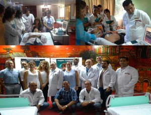
Curso_DTC_octubre_2018Entre los días 22 – 25 de octubre de 2018 sesionó en el Centro de Investigaciones Médico Quirúrgicas, organizado por el propio centro y la Organización Nacional de Trasplantes el Curso teórico práctico intensivo de neurosonología Doppler transcraneal, impartido por la Profesora Silvia Cocorullo de Argentina. Con el objetivo de “formar a formadores” para replicar el curso, participaron intensivistas de los hospitales Saturnino Lora (Santiago de Cuba), Vladimir I Lenin (Holguín), Manuel Ascunce (Camagüey), Arnaldo Millian (Villa Clara), Calixto García, Carlos J. Finlay y Juan Manuel Márquez (La Habana). El evento fue patrocinado por DWL.

0

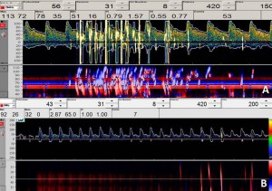
DTC_EmbEl caso A fue un paciente masculino que al ingreso en UCI se diagnosticó síndrome antifosfolipido y posterior al hallazgo del Doppler se realizó ecocardiografía que demostró trombo en AD y agujero oval permeable, que requirio de cirugia cardiaca. El caso B fue una paciente femenina con diagnóstico de cirrosis hepática por VHC. El hallazgo de émbolos tardíos nos hizo sospechar shunts intrapulmonares posiblemente asociados a síndrome hepato pulmonar.

0

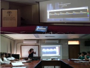
SonografiaDurante los días 8 y 9 de abril de 2016 sesionó el I Taller Nacional de Neuro Sonología y Hemodinámica Cerebral, organizado por el Servicio de Neurovascular del Instituto Nacional de Neurología y Neurocirugía. El evento contó con la participación de la destacada profesora argentina Silvia Cocorullo. El día 11 de abril, la profesora nos brindó en la UCI del CIMEQ, una excelente jornada teórico práctica sobre Sonografía Doppler transcraneal. En nuestro servicio se introdujo esta técnica en 2002. La primera publicación fue: Transcraneal Doppler sonography in brain death diagnosis (Restaurative Neurology and Neuroscience 21 (2003). Desde ese momento es parte de nuestras líneas de investigación con los temas encefalopatías hepática y asociada a sepsis. Sea este post el comienzo de una nueva etiqueta.

Filed under Uncategorized by on . Comment#

0

En este trabajo se caracterizo el patrón de flujo sanguíneo cerebral a través de Doppler transcraneal en un grupo de pacientes con fallo hepático fulminante. Conclusiones: El patrón mas frecuente fue el de hipoperfusión cerebral, con velocidades medias en arteria cerebral media disminuida e incrementos del índice de pulsatilidad.

Abdo A, et al: Transplantation Proceedings, 35, 1859–1860 (2003)